Fig. 2.

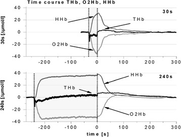
Typical kinetics of ΔO2Hb, ΔHHb, and ΔTHb during and after 30-s and 240-s cycling at 80 % in relation to baseline values (set to zero). Note the marked overshoot in ΔO2Hb following the 240-s exercise interval compared to the 30-s interval

Typical kinetics of ΔO2Hb, ΔHHb, and ΔTHb during and after 30-s and 240-s cycling at 80 % in relation to baseline values (set to zero). Note the marked overshoot in ΔO2Hb following the 240-s exercise interval compared to the 30-s interval