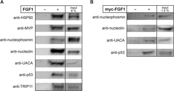
Figure 3.

Verification of intracellular FGF1 partners by co‐precipitation. A: BJ cell lysate was incubated with SBP‐FGF1 and then with dynabeads, or with dynabeads alone. Protein complexes were eluted from the beads with sample buffer, resolved by SDS‐PAGE and subjected to Western blotting using specific antibodies as indicated. B: HEK 293 cells transiently transfected with myc‐FGF1 were lysed and then subjected to immunoprecipitation followed by SDS‐PAGE and Western blotting with an anti‐nucleolin, anti‐p53, anti‐nucleophosmin, or anti‐UACA antibody. As controls non‐transfected cells were used.
