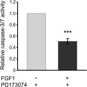
Figure 5.

FGFR‐independent anti‐apoptotic activity of exogenous FGF1. NIH3T3 cells were serum starved for 24 h and then treated for 6 h with FGF1 (200 ng/mL) and heparin (10 U/mL) in the presence of the specific FGFR tyrosine kinase inhibitor, 100 nM PD173084. Apoptosis was measured using ApoLive‐Glo Multiplex Assay (Promega) Next, caspase‐3/7 activity, reflecting the apoptosis progression, along with cell viability were measured using ApoLive‐Glo Multiplex Assay (Promega). Caspase‐3/7 activity data were divided by cell viability values, then normalized toward the cells untreated with FGF1 (control), and denoted as relative caspase‐3/7 activity. Graphs represent the mean ± SD of four independent experiments (***P < 0.001).
