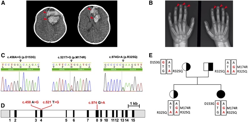
Figure 1.
Radiologic images of clinical phenotypes, sequencing results of GGCX genotypes, and pedigrees of the patient. (A) Computed tomography brain scan at hospital admission, showing a large area of bleeding on the left frontal lobe (arrowheads) and deviation of the midline. (B) Hand x-ray showing short distal phalanges of the fingers (arrowheads), a characteristic of Keutel syndrome. (C) Chromatograms of the identified sequence variations demonstrating heterozygous mutations in GGCX at c.458 A>G, c.521 T>G, and C.974 G>A. (D) The intron-exon organization of GGCX with the location of the mutations as identified in the patient. The 5′ and 3′ untranslated regions are shown in grayscale, the exons are indicated by black rectangles, and the introns are represented by clear rectangles. (E) The pedigrees of the patient’s family. Squares, male; circles, females; black-filled circle, disease-affected patient; black half-filled square/circles, carrier. Mutated nucleotides are in red; mutated amino acid residues are indicated on the side.

