Fig. 1.

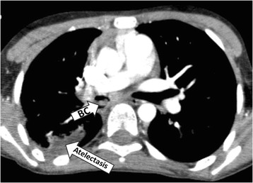
Thoracic computed tomography of case 1, revealing a solid 18 × 5 mm mass in the right main bronchus (BC) and post-obstructive atelectasis in the right upper lobe

Thoracic computed tomography of case 1, revealing a solid 18 × 5 mm mass in the right main bronchus (BC) and post-obstructive atelectasis in the right upper lobe