
Fig. 3.
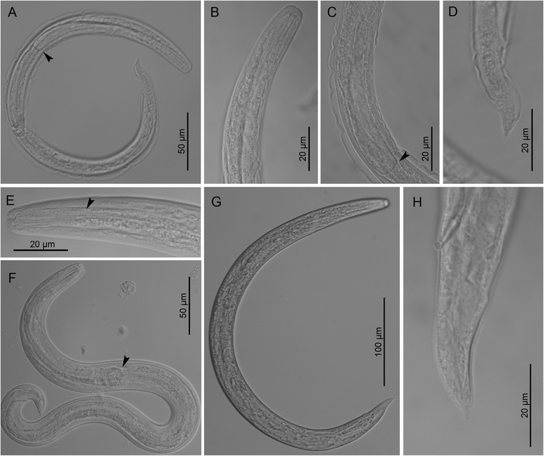
Third stage larvae of Crenosoma vulpis at different days post-infection (dpi), lateral view. Larva at 10 dpi, note the oesophago-intestinal junction (arrowhead) (a); Anterior extremity at 10 dpi (b); Posterior part of oesophagus at 10 dpi (c), note the oesophago-intestinal junction (arrowhead) and the loose cuticle of the second larval stage; Tail extremity at 10 dpi (d); Anterior extremity at 14 dpi (e), note the border between smooth and glandular parts of the oesophagus (arrowhead); Larva observed in vivo at 15 dpi (f), note the oesophago-intestinal junction (arrowhead); Larva at 150 dpi (g); Tail extremity at 180 dpi (h)