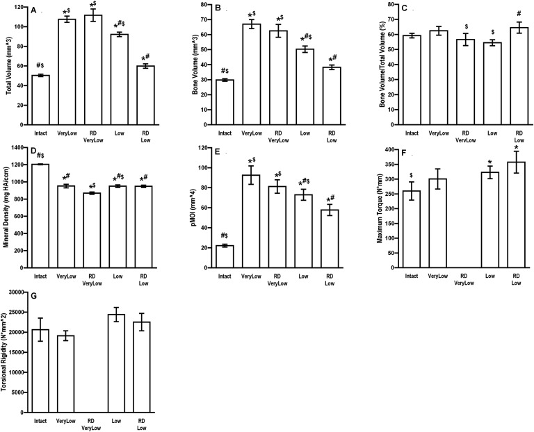
Fig. 8.
Physical properties of segmental defects treated with 5.5 μg of rhBMP-2 and reverse dynamization. Defects were stabilized initially with an external fixator of very low (25.4 N/mm) or low (114 N/mm) stiffness. After 2 weeks, the stiffness was increased to 254 N/mm (high stiffness) in the groups receiving reverse dynamization (RD Low and RDVeryLow). The animals treated with continuous (non-dynamized) fixation continued with low or very low-stiffness fixators (Low or VeryLow). After another 6 weeks, the femora were harvested and the defects were analyzed with μCT (Figs. 8-A through 8-E) and mechanical testing (Figs. 8-F and 8-G). The error bars indicate the standard error of the mean. #Significantly different from the RD very-low-stiffness group. *Significantly different from the intact, contralateral femur. $Significantly different from the RD low-stiffness group. Intact = contralateral femur, VeryLow = very-low-stiffness fixator (25.4 N/mm) throughout the entire 8 weeks, Low = low-stiffness fixator (114 N/mm) throughout the entire 8 weeks, RD VeryLow = very-low-stiffness fixator (25.4 N/mm) for the first 2 weeks followed by high-stiffness fixator (254 N/mm) for the remaining 6 weeks, RD Low = low-stiffness fixator (114 N/mm) for the first 2 weeks followed by high-stiffness fixator (254 N/mm) for the remaining 6 weeks, HA = hydroxyapatite, and pMOI = polar moment of inertia.

