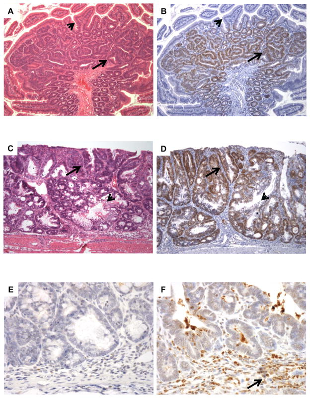
Figure 2. Id1 is overexpressed in mouse models of gastrointestinal neoplasia.
A, intestinal adenomas that develop in ApcMin/+ mice consist of crowded neoplastic crypts with mucin depletion (arrow) compared to the background small bowel mucosa (arrowhead). B, these tumors show strong Id1 staining (arrow), whereas the non-neoplastic epithelium is negative (arrowhead). C, Treatment with azoxymethane followed by several cycles of dextran sodium sulfate produced colonic epithelial neoplasia (arrow) comingled with non-neoplastic epithelium (arrowhead). D, colitis-associated neoplasia show strong, diffuse nuclear staining for Id1 (arrow) compared to entrapped non-neoplastic crypts (arrowhead). E, immunohistochemical stains for myeloperoxidase demonstrate a paucity of granulocytes in intestinal adenomas arising in ApcMin/+ mice, indicating that these lesions are not associated with inflammation. F, neoplasms associated with experimental colitis contain numerous inflammatory cells (arrow) that show strong staining for myeloperoxidase.

