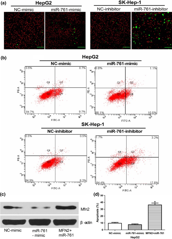
Figure 5.

MiR‐761 inhibitor affects mitochondrial function and MFN2 is involved in the MiR‐761 inhibitor‐mediated apoptosis. (a) Representative images show apoptosis and a decreased mitochondrial membrane potential (with enhanced green fluorescence and decreased red fluorescence) in SK‐Hep1 cells after transfection with the miR‐761 inhibitor (Scale bar: 50 μM.) (b) HepG2 and SK‐Hep1 cells were stained with a two‐color ROS detection kit and analyzed using flow cytometry after transfection with the miR‐761 inhibitor or miR‐761‐mimic. (c) Western blotting was used to detect Mfn2 expression in HepG2 cells after co‐transfection with miR‐761 and MFN2 plasmids lacking the 3′‐untranslated region (3′UTR) region; β‐actin was used as an internal control. (d) The apoptosis of HepG2 after co‐transfection with miR‐761 and MFN2 plasmids lacking the 3′UTR region. *P < 0.01.
