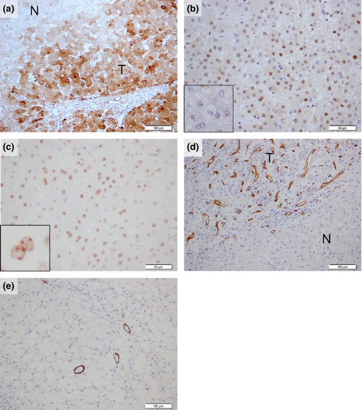
Figure 2.

Positive immunohistochemical expression pattern for each antibody. (a) CAP2 and (b) HSP70 showed a strong expression in the tumoral compared with the non‐tumoral liver ((b) inset) and (c) Bmi‐1‐positive regions showed clear “dot‐pattern” staining (inset). (d) Areas of CD34‐positive sinusoidal vascular architectural and (e) h‐caldesmon‐positive unpaired vessels were seen in high‐grade early hepatocellular carcinoma (HGeHCC). N, non‐tumoral liver; T, tumor. Scale bars = (a),(d),(e) 100 μm; (b,c) 50 μm.
