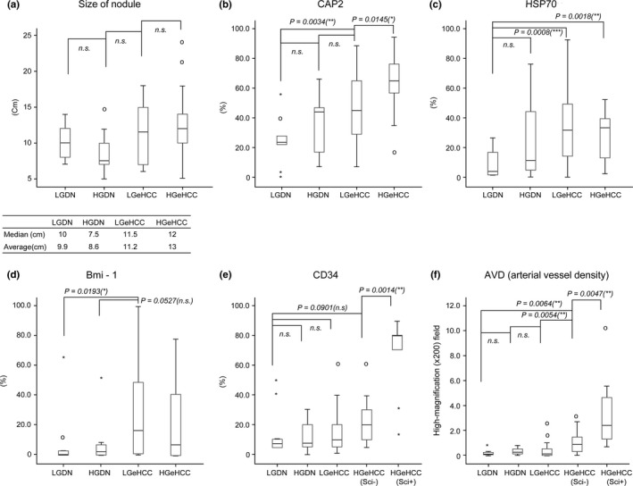
Figure 5.

Values of histopathological factors in each type of lesion. (a) Size of nodule; the immunohistochemical expression of (b) CAP2, (c) HSP70, (d) Bmi‐1 (e) and CD34; and (f) the arterial vessel density (AVD) were compared for each type of lesion. Nodule size, CAP2 expression, CD34 expression and AVD were increased for LGeHCC and/or high‐grade early hepatocellular carcinoma (HGeHCC) compared with those of DN. CAP2 expression and AVD in HGeHCC were statistically higher than those in low‐grade early hepatocellular carcinoma (LGeHCC) (Mann–Whitney U‐test). CD34 expression and AVD in scirrhous components (HGeHCC Sci+) were significantly higher than those in the non‐scirrhous components (HGeHCC Sci−) of HGeHCC (Mann–Whitney U‐test). Circles, outlying data. *P < 0.05, **P < 0.01, ***P < 0.001; n.s., not significant.
