
Figure 1.
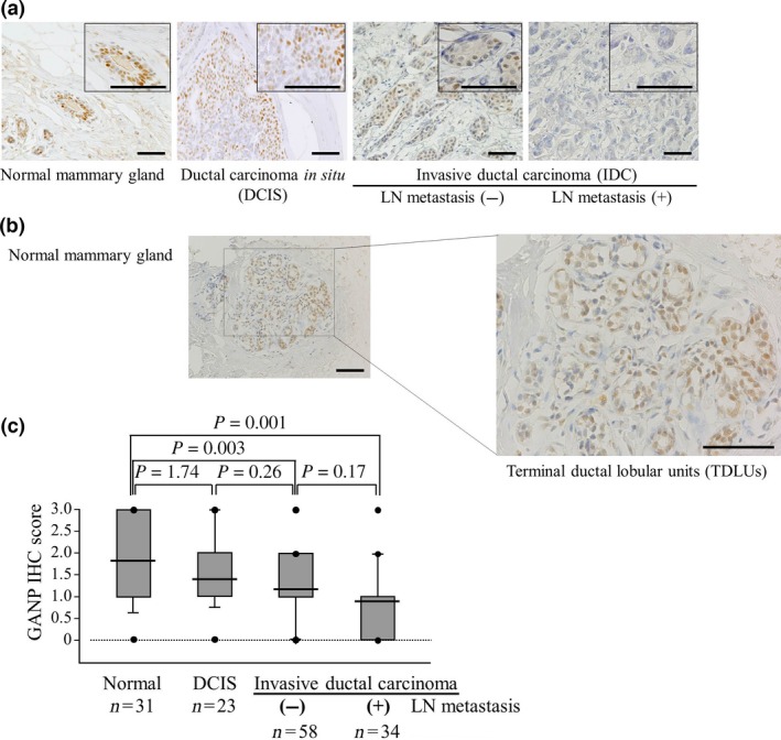
Reduced expression of GANP is significantly associated with the degree of malignancy in breast cancers. (A) Representative images of GANP immunostaining of a normal mammary gland, ductal carcinoma in situ and invasive ductal carcinoma. Images at higher magnification are shown in the insets. Scale bar = 100 μm. (B) GANP expression in terminal duct lobular units. Representative images of GANP immunostaining are shown. Scale bar = 200 μm. (C) A comparison of GANP expression among the four groups is shown by immunohistochemistry (IHC) scores using box plots with whiskers defining the range of scores. LN, lymph node.