
Figure 5.
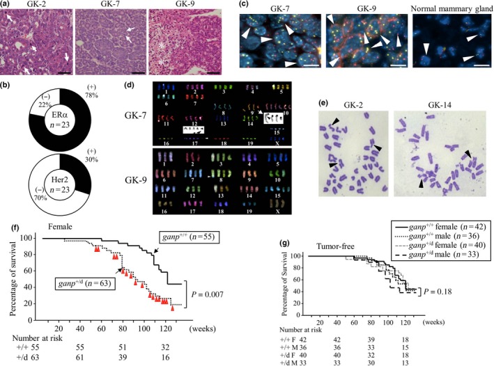
Female multiparous ganp +/d mice developed mammary gland tumors after aging. (A) Representative images of H&E staining of breast cancer tumors isolated from ganp +/d mice. Mitotic cells and necrotic lesions are indicated by arrows and asterisks, respectively. Scale bar = 100 μm. GK‐2, GK‐7, and GK‐9 are tumor specimens from different mice. (B) Expression levels of estrogen receptor α (ERα) and human epidermal growth factor receptor 2 (Her2) in the tumors. ERα and Her2 were examined by immunohistochemistry, and the frequencies of positive tumors are displayed in pie charts (n = 23). (C) Polyploidy in tumor sections. Dual centromere signals of mouse chromosome 14 (red) and 16 (yellow) were detected in the control mammary gland specimen (arrowheads). Two independent tumors (GK‐7 and GK‐9) showed more than two signals per cell (arrowheads). Scale bar = 20 μm. (D) Multicolor FISH analysis of cell lines. Abnormalities included increases in the numbers of chromosomes and translocations, as shown for chromosomes 9 and 17 (arrowheads in GK‐7). (E) Chromosome analysis of long‐term cultured tumor cell lines. Chromosomal breakages are shown (arrowheads). (F) Survival rates of female ganp +/+ (littermates) and ganp +/d mice. The life spans of the ganp +/d mice were shorter than those of the ganp +/+ mice (log–rank [Cox–Mantel] test). (G) Survival of individual tumor‐free mice was not significantly altered. Data were analyzed as in (F).