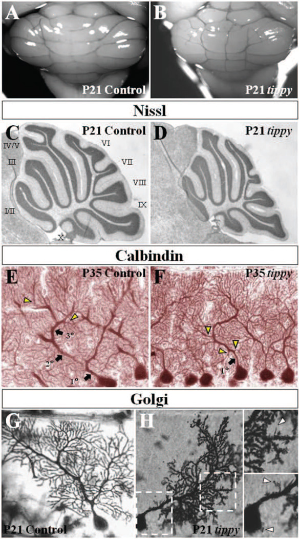
Figure 1. Abnormal mutant Purkinje cells without gross cerebellar dysmorphology in homozygous tippy mutant mice.
A,B, Whole cerebella of tippy mutant mice (B) exhibit grossly normal morphology with no significant differences from control cerebella (A). The midline structure in (B) is a prominent blood vessel in this particular specimen, a feature that is variable in both wild-type and mutant animals. There is no evidence of aberrant foliation or midline fusion deficits in tippy animals. C,D, Nissl stained mid-sagittal sections similarly demonstrate normal foliation pattern and cortical laminar structure in mutant (D) and control (C) mice. Numeric assignments for the lobules are shown for the control mice (C). E,F, Calbindin stained sagittal sections of cerebella from control (E) and tippy mutants (F) illustrate a dendritic branching malformation in mutant mice. In contrast to the stereotypical hierarchical Purkinje cell branching pattern displayed in control mice with spiny branchlets emerging only from tertiary branches (E, arrowheads), tippy mutant Purkinje cells display no clear secondary or tertiary shaft dendrites and spiny branchlets emerge directly from the primary dendrite (F, arrowheads). G,H, Golgi-stained Purkinje cells of control (G) and tippy mutant mice (H) reveal a loss of parasagittal planarity as well as a dendritic spine malformation with dendritic spines of immature morphology abnormally studding the proximal dendrite (H, top inset, arrowhead) and soma (H, bottom inset, arrowheads) in the mutant. Left and right boxed areas are shown at higher magnification as bottom and top insets, respectively.

