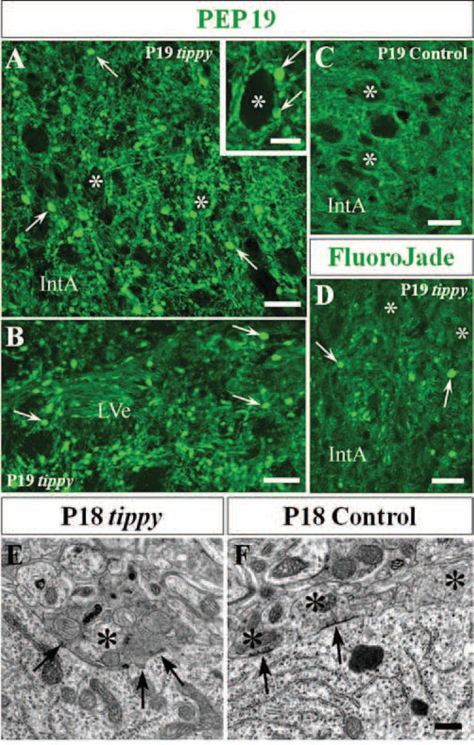
Figure 3. Degenerating Purkinje cell axons and terminals in cerebellar and vestibular nuclei of homozygous tippy mutant cerebella.
A–D, Tippy mutant cerebellar sections demonstrate abundant axonal spheroids (A,B,D, arrows) in the nucleus interpositus anterior (IntA) and the lateral vestibular nucleus (LVe) after staining with PEP 19 (A,B) or Fluoro-Jade C (D) while there are no spheroids present in the IntA of control cerebella (C). PEP19-positive spheroids (arrows) are apposed to the cell body of a large efferent neuron (asterisks) in the medial cerebellar nucleus (A, inset). E,F, Electromicrographs of Purkinje cell terminals in LVe of tippy mutant (E) and control (F) mice illustrate that the cell body of a normal, large efferent neuron in the LVe of control mice is surrounded by normal Purkinje axon terminals (F, asterisks), three of which contain dispersed small mitrochondia and pleomorphic synaptic vesicles. In contrast, the cell body of a large efferent neuron in the LVe of mutant mice is directly apposed by a swollen dystrophic terminal of a Purkinje cell axon (E, asterisk). The organelles in the dystrophic terminal closely resemble those observed in the terminal of the recurrent collateral in the cortex shown in Fig. 4F. Arrows indicate symmetric axo-somatic synapses. Scale bars: A–D, 20 µm; inset in A, 10 µm; E, F, 0.5 µm.

