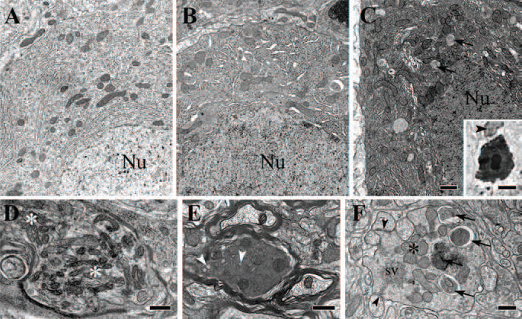
Figure 4. Electron microscopy of normal and degenerating Purkinje cell bodies and axons in P18 homozygous tippy mutant mice.
A–C, Illustration of the normal appearance of a Purkinje cell body (A), a dystrophic cell body with enlarged ER and an autophagosome (B), and a shrunken cell body in advanced stage of degeneration showing increased electron density of the cytoplasm, nucleoplasm and nuclear bodies, and a remarkable increase in lysosome-like and autophagosome-like bodies (C, arrows). A microglial phagocyte (arrowhead) approaching a shrunken, degenerated Purkinje cell body (C, inset) that is already surrounded by dilated Bergmann glial profiles (unlabeled white halo). D, A Purkinje cell axon forms a spheroid near the Ranvier heminode and contains clumped mitochondria (asterisks) interspersed with ER vesicles and small autophagosome-like bodies. E, A Purkinje cell axon in the folial white matter is undergoing dark degeneration. Arrowheads indicate vesiculated ER. F, A swollen terminal of a Purkinje cell recurrent axon collateral in the molecular layer shows advanced dystrophic signs, as indicated by assembled mitochondria (asterisk), autophagosomes (arrows), and clumped pleomorphic synaptic vesicles (sv). The degenerating profile forms symmetric junctions with Purkinje cell spines (arrowheads). Nu, cell nucleus; sv, synaptic vesicles. Scale bars: A–C, 1 µm; inset in C, 5 µm; D–F, 0.5 µm.

