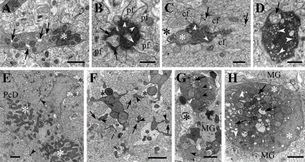
Figure 5. Electron microscopy of degenerating Purkinje cell dendrites in P19 homozygous tippy mutant mice.
A, A dendritic spiny branchlet contains moderately electron dense cytoplasm, abnormal dilated endoplasmic reticulum (arrows) and clumped mitochondria (asterisk). B, A degenerating spiny branchlet is surrounded by parallel fiber (PF) varicosities and appears shrunken. Two of its spine synapses (arrows) are still recognizable and PF synapses (arrowheads) are also present on the shaft, possibly corresponding to retracted spines. C, A degenerating medium-sized dendritic branch appears shrunken, contains an enlarged mitochondrion (white asterisk), is partially surrounded by dilated Bergmann glial processes (black asterisk) and forms synaptic junctions (arrowheads) with climbing fiber varicosities (CF) containing large dense core vesicles (arrows). D, An overtly degenerating second or third order Purkinje cell dendrite contains double membrane-bound, autophagosome-like bodies (arrow) and clusters of dilated or vesiculated ER cisterns (arrowheads). E, The main stem of a Purkinje cell dendrite (PcD) appears to undergo the initial stage of degeneration; the mitochondria—no longer separated by neurofilaments and microtubules—have formed two large assemblies (white asterisks); some of the ER cisterns appear dilated and the surrounding Bergmann glia profiles are swollen (black asterisk). Arrowheads point to synapses on the dendritic stem. F, Segmented Purkinje cell branchlets undergoing dark degeneration, are provided with spines (arrowheads) synapsing with parallel fibers (arrows). The branchlets contain small and compact clumps of enlarged mitochondria and are nearly encircled by dilated Bergmann glial processes (asterisks). G,H, Second and first order branches of a Purkinje cell dendrite undergoing dark degeneration contain enlarged mitochondria and autophagosome-like bodies, and are engulfed by Bergmann glial (black asterisk) and microglial (MG) cell processes. Cisterns of the endoplasmic reticulum (white arrowheads) have become vesiculated and centralized, separating from cytoskeletal elements (white asterisks). The mitochondrial matrix show varying density. Arrowheads indicate mitochodrial appositions and arrows point to autophagosomes. pf, parallel fiber; cf, climbing fiber; PcD, Purkinje cell dendrite; MG, microglia. Scale bars: A, E, G, H, 1 µm; B–D, F, 0.4 µm.

