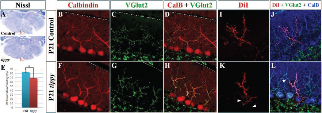
Figure 6. Increased climbing fiber terminals within a reduced dendritic domain and altered pattern of innervation in homozygous tippy mutant cerebella.
A, Nissl stained coronal sections demonstrate qualitatively normal inferior olivary neurons (IO) in both control and tippy mutant mice. B–L, However, confocal imaged sagittal sections of control (B–D) and tippy mutant (F–H) cerebella stained with calbindin (red; B,F) and VGluT2 (green; C,G) illustrate marked clustering of VGluT2-positive climbing fiber (CF) terminals (G) and increased density of CF terminals on the proximal dendrite and soma of tippy mutant PCs (H) compared to control (D). Quantitative analysis of CF innervation territory in P21 mice indicates a significant reduction in the domain encompassed by CF terminals in mutant mice compared to controls (E). n = 10 for WT, n = 11 for mutants. Further, confocal imaged sagittal sections of anterogradely DiI-labeled CFs (red), VGluT2-labeled CF terminals (green) and calbindin-labeled PCs (blue) in control (J) and tippy mutant (L) mice illustrate alterations in the pattern of CF innervation in mutant cerebella. Unlike single DiI-labeled CFs in control mice that travel a direct route to PCs (I), DiI-labeled CFs in the mutant cerebellum travel a circuitous path, intertwining with other DiI-labeled CFs in the PC layer (K, arrowheads). Triple-labeled images (J,L) highlight the increased thickness of CFs in the mutant, presence of DiI-labeled, VGluT2-positive puncta on the mutant PC soma and proximal dendritic shaft as well as the presence of of VGluT2-positive, DiIunlabeled terminals on the same PC (L, arrowhead) as VGluT2-positive, DiI-labeled puncta, suggesting multiple innervation. Data are expressed as mean ± SEM. *p < 0.00001, Student’s t-test. The pial surface is indicated by the dotted line.

