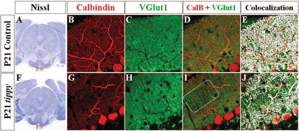
Figure 8. Decreased PF-PC synaptic contacts in homozygous tippy mutant mice.
A,F, Nissl stained coronal sections demonstrate qualitatively normal pontine nuclei in control (A) and tippy mutant (F) mice at P21. B–J, Confocal imaged sagittal sections stained with calbindin (red; B,G) and VGluT1 (green; C,H) likewise illustrate VGluT1-positive PF terminals in tippy mutant mice (H) distributed within an innervation domain and at density comparable to that seen in control cerebella (C). However, in contrast to the homogenous overlay between calbindin-labeled PC processes and VGluT1-labeled PF terminals seen in control mice (D,E), patches of PF terminals are seen without associated PC processes (I,J boxed regions) in tippy mutants. Colocalization of calbindin and VGluT1-labeled puncta are shown in white in (E,J).

