Summary
Elf and colleagues employed an elegant series of functional and biochemical assays to investigate the molecular mechanism of mutant calreticulin (CALR)-driven cellular transformation in myeloproliferative neoplasms (MPN). Mutant CALR is sufficient to induce MPN in mouse transplantation experiments and transformation is dependent upon physical interaction mediated by the positive electrostatic charge of the mutant CALR C-terminal domain and the thrombopoietin receptor (MPL) (1).
Myeloproliferative neoplasms (MPN) are a group of clonal hematopoietic stem cell malignancies that give rise to discrete hematological diseases (2). BCR-ABL negative MPNs comprise a group of molecularly related but phenotypically distinct myeloid malignancies that include polycythemia vera (PV), essential thrombocythymia (ET), and primary myelofibrosis (PMF). Patients with PV present clinically with predominant erythrocytosis and myeloid hyperplasia; and patients with ET or PMF present with predominant megakaryocytic hyperplasia and either thrombocytosis or bone marrow fibrosis, respectively. BCR-ABL negative MPN are united by aberrant cytokine signaling caused by recurrent oncogenic driver mutations that lead to downstream JAK-STAT activation and cellular transformation. The most common driver mutation in MPN is JAK2V617F which is present in >95% of PV cases, and 50–60% of ET and PMF cases (2). Additional driver mutations include activating mutations in MPL in 3–7% of cases of ET and PMF (2–4) and less commonly, deactivating mutations in negative regulators of JAK-STAT signaling (5).
Recently, in an attempt to identify additional driver mutations in MPN, whole exome sequencing of ET and PMF patients wild-type for both JAK2 and MPL (double-negative patients) identified mutually exclusive CALR mutations in 67% of ET patients and 88% of PMF patients with double-negative disease (6, 7). To date, 36 different insertion or deletion (indels) CALR mutants have been identified (6). All mutations are located within exon 9 and cause a one base-pair (+1 bp) reading frameshift mutation that generates a novel C-terminal peptide (6, 7).
The wild-type CALR gene is highly conserved and encodes a 417 amino acid multifunctional protein that localizes predominately to the lumen of the endoplasmatic reticulum (ER), where it functions as a protein-folding chaperone and a Ca2+ storage molecule (8). CALR is made up of three protein domains: the N-terminal (N) and proline-rich (P) domains are primarily responsible for most of the protein folding functions, whereas the C-terminal domain binds to Ca2+ with a series of negatively charged amino acids and has an ER retention signal (KDEL). Notably, the novel C-terminal domain found in mutant CALR MPN patients lacks the KDEL ER retention motif and codes for a series of positively charged amino acids. CALR has never been found to be mutated in cancer prior to its identification in MPN and this has prompted several important questions concerning the role of mutant CALR in the pathogenesis of these diseases: Can mutant CALR alone drive oncogenic transformation? What is the mechanism by which the CALR mutant C-terminus is involved in disease pathogenesis? Why are CALR mutations exclusive from JAK2V617F mutations?
To investigate whether mutant CALR is capable of inducing an MPN phenotype in mice, Elf and colleagues transduced wild-type c-Kit-enriched primary bone marrow cells with retroviral vectors expressing either empty vector, wild-type human CALR cDNA (CALRWT) or a human mutant CALR cDNA (CALRMUT) (the most commonly found CALR mutation P.L367fs*46; results in a 52 bp deletion) and transplanted these cells into lethally irradiated congenic recipient mice. 16 weeks post-transplantation, CALRMUT recipient mice developed an ET-like phenotype including thrombocytosis and megakaryocytic hyperplasia suggesting specific involvement of the megakaryocytic lineage. Furthermore, analysis of CALR expression in primary bone marrow cells from CALR-mutant MPN patients showed high expression in the megakaryocyte lineage and in immature myeloid cells and was largely absent from erythroid or mature myeloid lineages.
The megakaryocyte lineage-specific phenotype observed in CALRMUT transplantation experiments and the megakaryocytic lineage restricted expression of CALR in patients prompted the authors to look at the potential role of MPL in CALRMUT transformation. Using an established cellular transformation assay, first pioneered by Daley et al (9), Elf and colleagues show that CALRMUT expressing Ba/F3 murine hematopoietic cells could not convert to IL-3 independence alone and instead required co-expression of MPL but not other hematopoietic cytokine receptors including the granulocyte-colony stimulating factor receptor (G-CSFR) or the erythropoietin receptor (EPOR). This effect was reproduced in UT-7 megakaryocytic cells in which expression of CALRMUT but not CALRWT allowed for GM-CSF independent growth with MPL co-expression.
Given that the results of the cellular transformation assay were obtained with ectopic expression of CALR variants, the authors recapitulated the transforming ability of mutant Calr under endogenous expression levels. CRISPR/Cas9 gene editing technology was used to generate a +1 bp frameshift mutation in exon 9 of murine Calr. Ba/F3-MPL cells stably expressing Cas9 were infected individually with two distinct small guide RNAs (sgRNA), which led to IL-3-independent growth that was not observed in Calr-targeted parental Ba/F3-Cas9, or Calr-targeted Ba/F3-Cas9 cells overexpressing each EPOR or G-CSFR. Sequencing of genomic DNA from these experiments revealed that each distinct sgRNA was able to generate indels that led to a +1 bp frameshift mutation. Together these data provide definitive evidence that generation of a heterozygous 1 bp frameshift mutation (as seen in human MPN (6, 7)) in the endogenous Calr locus coupled with MPL expression is sufficient for cellular transformation.
RNA sequencing of CALRMUT transformed Ba/F3-MPL cells showed significant enrichment for Stat3 and Stat5 gene signatures and western blotting confirmed activation of the JAK-STAT signaling pathway. Notably, cellular transformation and JAK-STAT activation was not observed in parental Ba/F3, Ba/F3-EPOR or Ba/F3-G-CSFR expressing CALRMUT. Furthermore, knockdown of Jak2 by shRNA led to a significant decrease in proliferation of Ba/F3-MPL cells with CALRMUT and treatment with the JAK inhibitor ruxolitinib abolished downstream Stat3 activation. This suggests that mutant CALR is dependent on MPL for transformation and that co-expression leads to downstream JAK-STAT signaling, which can be abrogated by targeting JAK2.
To study the structural and functional basis of oncogenic transformation Elf et al generated individual CALR domain mutants. They found that ectopic expression of either the wild type N, P, or C domains alone, or the mutant CALR C-terminal domain alone was not sufficient to confer IL-3 independent growth to Ba/F3-MPL cells, suggesting that the CALR mutant C-terminal domain is necessary but alone is not sufficient to transform cells. Furthermore, removal of the C-terminal KDEL sequence alone did not lead to cellular transformation.
Next, Elf et al created a series of C-terminal deletion mutants in which 8–10 amino acids were individually deleted. All four mutants led to oncogenic transformation and equal outgrowth of Ba/F3-MPL cells suggesting that the transforming capabilities of CALRMUT are not due to specific residues within the C-terminus but rather are a shared property of the mutant C-terminal tail. CALR mutations in MPN patients encode for a positively charged C-terminus with a series of lysine (K) and arginine (R) residues that replace this normally negatively charged region (6). Strikingly, a C-terminal CALR domain mutant in which each K and R residue was substituted with neutral glycine (G) residues (CALRMUT-neutral) was no longer capable of transforming Ba/F3-MPL cells, while a control mutant in which each non-K and non-R residue was substituted with G (CALRMUT-positive) retained its transforming ability. Collectively, these data revealed an absolute requirement for MPL expression and for the positively charged C-terminal domain of mutant CALR for oncogenic transformation, which suggested a direct interaction between the two molecules. Elf et al then used FLAG tagged CALR variant immunoprecipitation experiments to definitively show that CALRMUT, but not CALRWT, physically interacts with MPL. Importantly, these results were recapitulated in CRISPR-targeted Ba/F3-MPL-Cas9 cells expressing pathophysiologically relevant levels of mutant Calr showing that this differential binding is not due to over-expression of mutant CALR. Finally, the authors showed that the positively charged amino acids of the mutant CALR C-terminal domain are required for physical interaction with MPL.
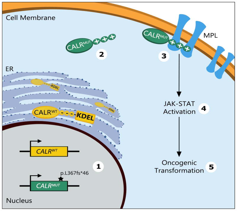
In summary, through extensive mutagenesis-based structure-function experiments and biochemical assays Elf et al provide a detailed mechanism for mutant CALR-mediated oncogenic transformation and offer explanations to many aspects of mutant CALR-mediated MPN (Figure 1). Specifically, this study shows that mutant CALR is sufficient to initiate an ET-like phenotype in vivo; that direct physical interaction between mutant CALR and MPL is essential for mutant CALR-mediated transformation and that this interaction is dependent upon the positive electrostatic charge of the mutant CALR C-terminus.
Figure 1. Schematics of the mechanism of mutant CALR-mediated oncogenic transformation.
(1) Frameshift mutations (here: p.L367fs*46) in exon 9 of the calreticulin (CALR) gene codes for mutant CALR (CALRMUT) with a positively charged C-terminal domain instead of the negatively charged C-terminus of wild-type CALR (CALRWT) protein. (2) CALRMUT also lacks the C-terminal endoplasmic reticulum (ER) retention signal (KDEL) present in CALRWT protein. (3) The positive charge of the CALRMUT C-terminus is required for direct binding to the thrombopoietin receptor (MPL). (4),(5) Binding of CALRMUT to MPL is required for downstream JAK-STAT activation and oncogenic transformation. Illustration by Barbara Walter.
The observation that mutant CALR activates downstream JAK-STAT signaling provides a biological basis as to why JAK2 and CALR mutations are mutually exclusive in patients with MPN and offers an explanation as to why CALR mutated patients respond to JAK inhibition. The study by Elf et al solidify JAK-STAT activation as the central pathway driving oncogenic transformation in MPN. Despite improvements in understanding the initiation of this process, JAK inhibition does not significantly alter the course of disease, highlighting the urgent need for novel disease-modifying drugs (2). Importantly, the mechanistic findings by Elf and colleagues provide a biological rationale to develop novel therapeutic inhibitors targeting the required physical interaction between mutant CALR and MPL in mutant CALR-mediated MPN.
Acknowledgments
R.F.S. has been supported by an MSTP training grant (T32GM007288), an individual NHLBI Fellowship (F30HL122085), and the NIH Cellular and Molecular Biology and Genetics Training Grant (T32GM007491). U.S. is the Diane and Arthur B. Belfer Scholar in Cancer Research of the Albert Einstein College of Medicine, and is a Research Scholar of the Leukemia & Lymphoma Society.
Footnotes
The authors declare that they do not have any relevant conflicts of interest.
References
- 1.Elf S, AN, Chen E, Perales-Paton J, Rosen EA, Ko A, et al. Mutant calreticulin requires both its mutant C-terminus and the thrombopoietin receptor for oncogenic transformation. Cancer Discov. 2016 doi: 10.1158/2159-8290.CD-15-1434. [DOI] [PMC free article] [PubMed] [Google Scholar]
- 2.Tefferi A. Myeloproliferative neoplasms: A decade of discoveries and treatment advances. American journal of hematology. 2016;91:50–8. doi: 10.1002/ajh.24221. [DOI] [PubMed] [Google Scholar]
- 3.Pikman Y, Lee BH, Mercher T, McDowell E, Ebert BL, Gozo M, et al. MPLW515L is a novel somatic activating mutation in myelofibrosis with myeloid metaplasia. PLoS Med. 2006;3:e270. doi: 10.1371/journal.pmed.0030270. [DOI] [PMC free article] [PubMed] [Google Scholar]
- 4.Pardanani AD, Levine RL, Lasho T, Pikman Y, Mesa RA, Wadleigh M, et al. MPL515 mutations in myeloproliferative and other myeloid disorders: a study of 1182 patients. Blood. 2006;108:3472–6. doi: 10.1182/blood-2006-04-018879. [DOI] [PubMed] [Google Scholar]
- 5.Tefferi A, Pardanani A. Myeloproliferative Neoplasms: A Contemporary Review. JAMA Oncology. 2015;1:97–105. doi: 10.1001/jamaoncol.2015.89. [DOI] [PubMed] [Google Scholar]
- 6.Klampfl T, Gisslinger H, Harutyunyan AS, Nivarthi H, Rumi E, Milosevic JD, et al. Somatic mutations of calreticulin in myeloproliferative neoplasms. N Engl J Med. 2013;369:2379–90. doi: 10.1056/NEJMoa1311347. [DOI] [PubMed] [Google Scholar]
- 7.Nangalia J, Massie CE, Baxter EJ, Nice FL, Gundem G, Wedge DC, et al. Somatic CALR mutations in myeloproliferative neoplasms with nonmutated JAK2. N Engl J Med. 2013;369:2391–405. doi: 10.1056/NEJMoa1312542. [DOI] [PMC free article] [PubMed] [Google Scholar]
- 8.Lu Y-C, Weng W-C, Lee H. Functional roles of calreticulin in cancer biology. Biomed Res Int. 2015:526524. doi: 10.1155/2015/526524. [DOI] [PMC free article] [PubMed] [Google Scholar]
- 9.Daley GQ, Baltimore D. Transformation of an interleukin 3-dependent hematopoietic cell line by the chronic myelogenous leukemia-specific P210bcr/abl protein. Proceedings of the National Academy of Sciences. 1988;85:9312–6. doi: 10.1073/pnas.85.23.9312. [DOI] [PMC free article] [PubMed] [Google Scholar]


