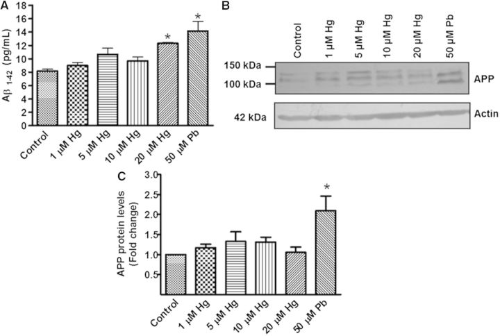
FIG. 3.
Effect of metals in Aβ-42 and APP levels. Differentiated SH-SY5Y cells (1.5 × 10⁶) were incubated with 1, 5, 10, or 20 µM of Hg or 50 µM of Pb for 48 h. Cells treated with 50 µM Pb were used as a positive control. The group without treatment was used as a control. A, Aβ-42 levels in total protein extracts. B, A representative blot of Hg and Pb effects on APP levels. C, Densitometric analysis of APP protein levels from 3 independent experiments; mean ± SE. Actin was used as a loading control. *P ≤ .05 different with respect to control cells according to 1-way ANOVA and Dunnet’s multiple comparison post-hoc tests. RA = retinoic acid.

