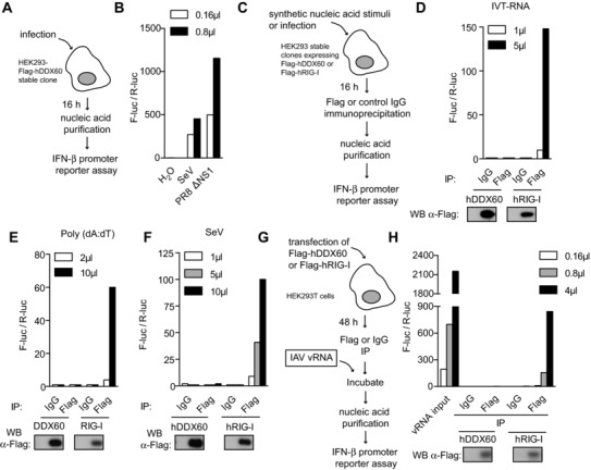
Figure 7.

IP of DDX60 does not coprecipitate stimulatory RNA. (A) Diagram of experiment conducted in (B) where the stimulatory capacity of nucleic acid purified from stable HEK293‐Flag‐hDDX60 cells infected with SeV or PR8ΔNS1 (MOI 1) was assessed by IFN‐β promoter reporter assay. Water was used as a control. (C) Diagram of experiment conducted in (D, E, and F) where Flag‐hDDX60‐stable HEK293 cells were transfected with (D) IVT‐RNA or (E) poly(dA:dT) or infected with (F) SeV. Sixteen hours later, Flag‐hDDX60 was immunoprecipitated with an anti‐Flag antibody. An isotype‐matched IgG IP was included as control. RNA was then purified from precipitates and stimulatory capacity was assessed by IFN‐β promoter reporter assay (above). IP samples were also tested for Flag expression by Western blot (below). Experiment was conducted in conjunction with HEK293‐Flag‐hRIG‐I cells. (G) Diagram of experiment conducted in (H) where HEK293T cells were transfected with Flag‐hDDX60. Forty‐eight hours later, Flag‐hDDX60 was immunoprecipitated with an anti‐Flag antibody and an isotype‐matched IgG IP was included as control. Flag‐hDDX60‐coated IP beads as well as control beads were incubated with RNA isolated from IAV (IAV vRNA). Following extensive washes, RNA was purified from precipitates and stimulatory capacity was assessed by IFN‐β promoter reporter assay. IP samples were tested for Flag expression by Western blot. The experiment was conducted in conjunction with Flag‐hRIG‐I transfection. For all experiments one representative of three independent experimental repeats is shown.
