
Figure 3.
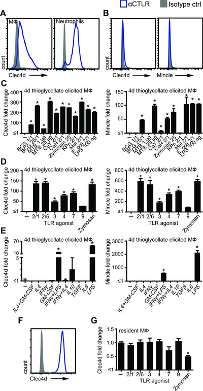
Modulation of Clec4d and Mincle expression by microbial stimuli. (A–F) Flow cytometric analysis of receptor expression on mouse peritoneal cells. (A) Peritoneal recruited cells 4 days after i.p. thioglycollate injection were analyzed for Clec4d expression (representative of >3 mice from two experiments). Plated thioglycollate‐elicited peritoneal macrophages were analyzed for Clec4d and Mincle expression under (B) resting conditions, which indicates the baseline for further stimulations (representative of >3 experiments) or after stimulation for 16 h with (C) microbial agents: BCG, M. bovis BCG; MTB, M. tuberculosis; CaY, C. albicans yeast; CaH, C. albicans hyphae; KPn, K. pneumoniae; Mal, Malassezia (representative of a single experiment), (D) TLR agonists (pooled data from two experiments performed in duplicates) or (E) cytokines (representative of a single experiment performed in duplicates). (F) Resident peritoneal cells were plated and (G) stimulated with TLR agonists for 16 h followed by Clec4d‐expression analysis by flow cytometry (pooled data from two experiments performed in duplicates). All data shown as mean + SD for single experiments and mean + SEM for pooled experiments of the indicate number of samples. Statistical significance was determined by ANOVA and Dunnett's multiple‐comparison posttests; *p < 0.05.