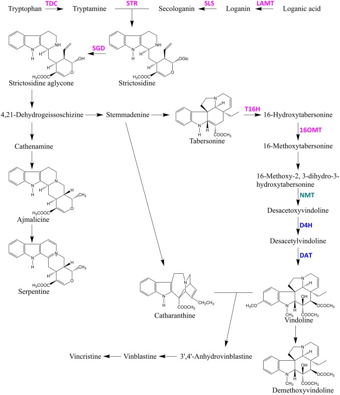
Fig. 1.
TIA metabolic pathway in Catharanthus roseus. Purple font represents TIA enzymes localized in ECs. Green represents the TIA enzyme localized in PCs. Blue represents TIA enzymes localized in ICs and LCs. D4H, desacetoxyvindoline 4-hydroxylase; DAT, deacetylvindoline 4-Oacetyltransferase; LAMT, loganic acid O-methyltransferase; NMT, 16-methoxy-2,3-dihydro-3-hydroxy-tabersonine N-methyltransferase; SGD, strictosidine β-glucosidase; SLS, secologanin synthase; STR, strictosidine synthase; T16H, tabersonine 16-hydroxylase; TDC, tryptophan decarboxylase; 16OMT, 16-hydroxytabersonine O-methyltransferase.

