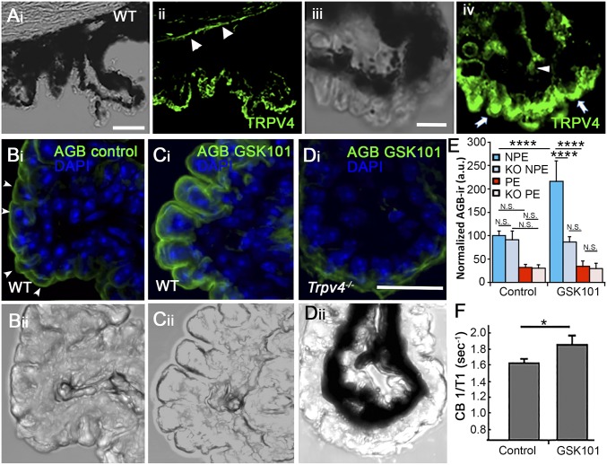
Fig. 1.
Localization and functional expression of TRPV4 in the mouse CB. (A, i and ii) Vertical cryosections of WT mouse retinas immunolabeled for TRPV4 show preferential localization to the basolateral layer corresponding to NPE, with additional signals in the limbal corneal epithelium (arrowheads). (i) Transmitted image of the CB and the supporting cornea; (ii) TRPV4 immunoreactivity. (Scale bar, 100 μm.) (iii and iv) Close-up of the CB epithelium, showing TRPV4-ir in putative NPE cells (arrows). Modest immunofluorescence is detected in the putative stromal area (arrowhead). (Scale bar, 20 μm.) (B–E) Excitation mapping shows TRPV4-evoked cation influx into CB ex vivo. CBs were fixed, stained with an anti-AGB antibody (FITC), and counterstained with DAPI (blue). ROIs of equal size were placed around DAPI-positive cells in the NPE layer and PE layer, respectively. (B, i and ii) Transmitted + fluorescence image of isolated unstimulated (PBS-treated) nonpigmented CB tissue. (C, i and ii) Transmitted + fluorescence image of nonpigmented CB tissue stimulated with GSK101 (100 nM) and tested for AGB-ir + DAPI. (D, i and ii) Transmitted + fluorescence image of Trpv4−/− CB tissue stimulated with GSK101 simultaneously with preparations in B and C. (Scale bar, 20 μm.) (E) Quantification of fluorescence from GSK101-treated vs. PBS-treated WT and KO sections. P < 0.005. (F) In vivo CB signal evaluated using MEMRI. Quantification of average CB 1/T1 from vehicle (n = 5) and GSK101-treated (100 μM; n = 5) eyes. Values are presented as means ± SEM; *P = 0.02, ***P < 0.005, ****P < 0.0001.

