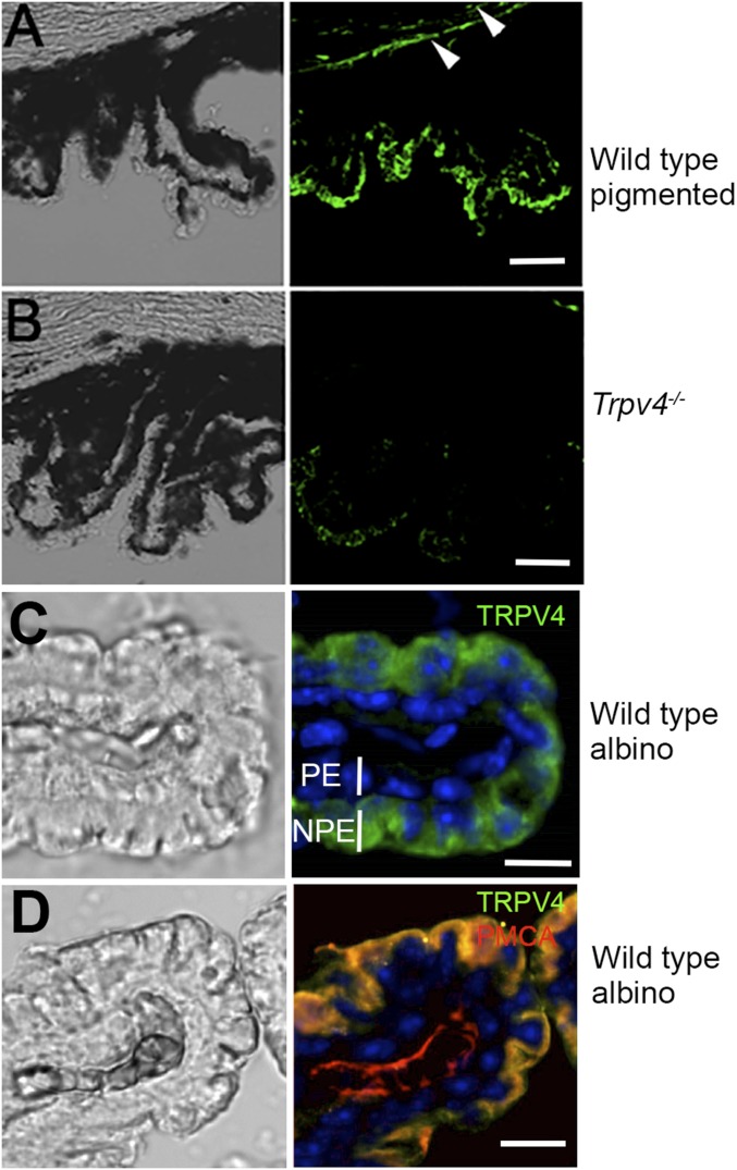
Fig. S1.
(A and B) Vertical sections of concomitantly labeled pigmented WT and Trpv4−/− ciliary bodies (see also Fig. 1A, ii and iv). (Scale bars, 100 μm.) The KO tissue exhibits little TRPV4-ir. Arrowheads in A point at TRPV4-ir in the corneal endothelium. (C) Nonpigmented CB tissue labeled for TRPV4 and counterstained for DAPI shows immunoreactivity predominantly in the NPE layer. (Scale bar, 20 μm.) (D) Nonpigmented CB. Double labeling with the anti-TRPV4 and anti-PMCA antibodies shows colocalization between TRPV4 and the calcium pump within the NPE plasma membrane. PMCA is also expressed in the stroma. (Scale bar, 20 μm.)

