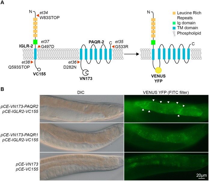
Fig 2. Novel alleles of PAQR-2 and IGLR-2, and interaction of IGLR-2 with PAQR-2.
(A) Schematic structures of the IGLR-2 and PAQR-2 proteins, with novel mutations indicated by red arrowheads. The VC155 and VN173 fragments added to the C and N terminal ends of IGLR-2 and PAQR-2, respectively, allows reconstitution of a full and fluorescent VENUS YFP protein if the two proteins come into close proximity. (B) Result of the BiFC experiment showing that IGLR-2 and PAQR-2 contact each other on cellular membranes. The top two panels show a transgenic worm co-expressing the fusion proteins depicted in (A); note the clear membrane-localized fluorescence indicative of IGLR-2 and PAQR-2 interaction. The middle two panels show a transgenic worm co-expressing the tagged IGLR-2 and a tagged PAQR-1 protein; note that only autofluorescent gut granules emit a signal, indicating that IGLR-2 and PAQR-1 do not interact with each other. The bottom two panels show a transgenic animal carrying the two empty vectors used in the BiFC experiments; note again that only autofluorescent gut granules emit a signal.

