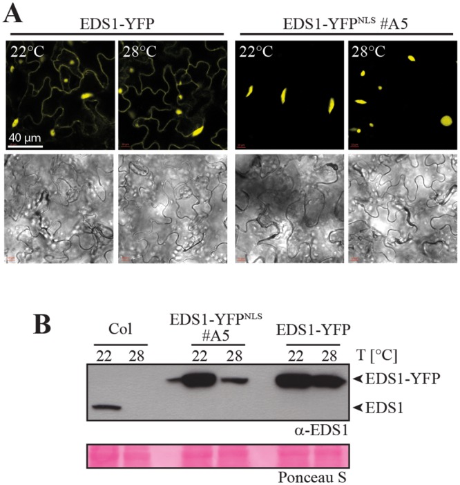
Fig 3. Temperature modulation of EDS1 protein accumulation and localization.

A. Confocal live cell images of representative leaf epidermal cells from EDS1-YFP and EDS1-YFPNLS #A5 plants grown at 22°C or 28°C. B. Immunoblot analysis of total protein extracts from 4-week-old plants grown at 22°C or 28°C separated by SDS-PAGE and probed with α-EDS1 antibody. Ponceau S staining of the membrane is shown as loading control.
