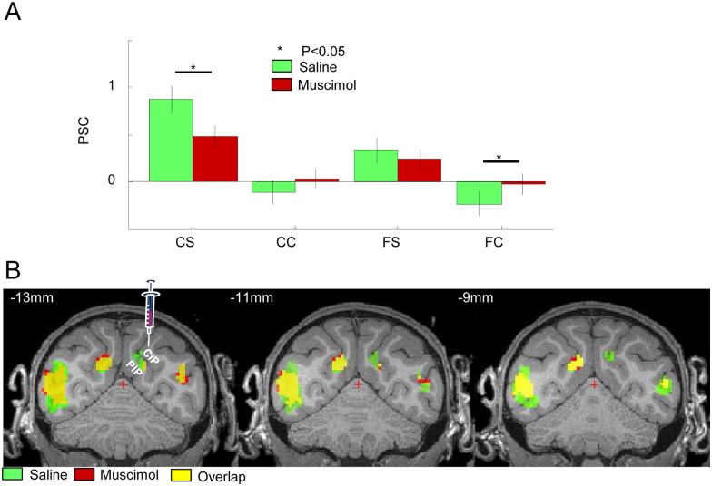
Fig 2. Effect of muscimol injection in CIP on the fMRI activations in the caudal IPS.
A. Group average percent signal change (compared to fixation) in the four experimental conditions. CS: curved stereo, CC: curved control, FS: flat stereo, FC: flat control. Green bars indicate saline sessions; red bars indicate muscimol sessions. * = p < 0.05. Raw values in [27]: cIPSr. B. Group average of the contrast [(curved stereo–curved control)–(flat stereo–flat control)] in the caudal IPS during saline and muscimol sessions (at p < 0.05 FWE corrected for multiple comparisons), plotted on coronal sections of the M12 anatomical template (average of three monkeys). Green: saline sessions, red: muscimol sessions, yellow: overlap between saline and muscimol sessions. Green voxels indicate significant depth structure activations during saline sessions but not during muscimol sessions, hence voxels that were affected by CIP inactivation. Red voxels indicate significant activations during muscimol sessions but not during saline session (mainly in the contralateral hemisphere). The left panel indicates the approximate location of the muscimol injection (see also S1B Fig). t values in [27]: stereo_sal.img/hdr and stereo_mus.img/hdr.

