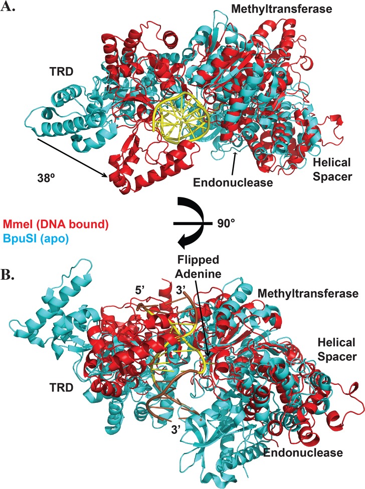
Fig 2. Structural comparison between MmeI and BpuSI.
(A) MmeI (red) bound to DNA (yellow) superimposed on apo BpuSI (cyan). The helicase connector, methyltransferase, and DNA target recognition domain (TRD) labels correspond to both structures, while the endonuclease domain is only visible in the BpuSI structure. A comparison of the two structures reveals an ~38° rotation in the TRD, which clamps down on the DNA to make specific contacts. The TRD as a whole shifts by ~27 Å between the two structures. (B) A 90° rotation of the view in (a) to show the relative position of the endonuclease domain.

