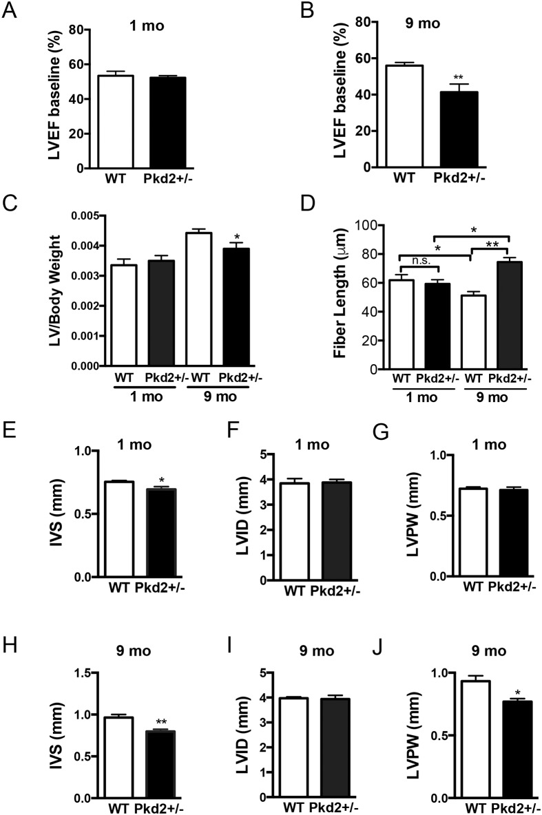
Fig 2. 9 mo Pkd2+/- mice have decreased ejection fraction and ventricular remodeling.
(A) 1 mo WT and Pkd2+/- mice have similar left ventricular ejection fraction values. (B) 9 mo Pkd2+/- mice have significantly lower left ventricular ejection fraction values compared with WT mice. (C) Left ventricular to body weight ratios were significantly reduced in 9 mo mice but were unchanged in 1 mo mice. (D) Cardiomyocyte lengths were significantly longer in 9 mo mice but are unchanged in 1 mo mice. (E-G) 1 mo Pkd2+/- mice have significantly thinner inner septum measurements compared with WT mice, but the posterior wall and interior diameter are the same. (H-J) 9 mo Pkd2+/- mice have significantly thinner left ventricular wall and inner septum measurements compared with WT mice. Data in A-C, E-J are representative of five 1 mo mice in each group, and eight and nine WT and Pkd2+/- 9 mo mice, respectively. Data in D are representative of at least two different animals per group with 22–45 cells measured from each animal. *p < 0.05, **p < 0.01, n.s. = not significant.

