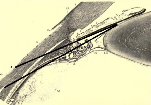
Figure 10.

The ciliary region of the eye.37
Notes: Added lines show the possibility of a couching needle passing through the sclera (as far posteriorly as the rectus muscle insertion), the ciliary body, the posterior chamber, and the iris–lens channel, while remaining posterior to the iris and anterior to the vitreous. The absolute limbus to rectus muscle distance, and the ciliary body length, are both 20%–23% less in the medial region, depicted here, compared with the lateral region,56 which is involved with the couching technique of Celsus, pseudo-Galen, and Paulus Aegineta. Nonetheless, the relative length of internal and external structures is unchanged.
Abbreviations: b, border of Bowman’s membrane; qB, vitreous base; Cs, corneoscleral limbus; Co, conjunctiva sclerae; d, Descemet’s membrane; Es, episcleral tissue; f, contraction furrows; vG, anterior border of the vitreous; hG, posterior border of the vitreous; Gk, vitreous nucleus; k1, ciliary and k2 pupillary crypts; Lhc, ligamentum hyaloideo-capsulare; Mr, medial rectus tendon; iZ, innermost; cZ, circular; vZ, anterior; qZ, middle; hZ, posterior zonula fibers; Z, zonular cleft.
