Figure 1.
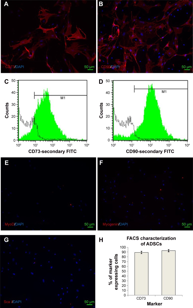
Characterization of rat adipose-derived stem cells (ADSCs) in culture.
Notes: ADSCs were cultured after isolation and cells at passage 2 were either seeded on glass coverslips and cultured in proliferation media in an undifferentiated state for 3 days or processed for FACS analysis. Expression of standard surface markers for ADSCs was confirmed by staining of CD73 (A) and CD90 (B). C and D depict percentage of CD73 and CD90 cells by FACS analysis. Absence of any contaminating muscle progenitor cells in ADSC cultures was revealed by the absence of IF staining for nuclear muscle markers MyoD (E), myogenin (F), and a nonspecific ADSC surface marker Sca I (G). FACS analysis (n=3) estimated that 89.42%±3.2% of cells expressed CD73, whereas CD90 was expressed by 92.80%±2.4% cells (H). FACS analysis also confirmed the negative staining of muscle markers (Pax7, MyoD, and myogenin) and absence of nonspecific surface marker (Sca I) in ADSCs. M1, represents gate used to define the positive cell population. Data is presented as mean ± SD.
Abbreviations: FACS, fluorescence-activated cell sorting; FITC, fluorescein isothiocyanate; DAPI, 4′, 6-diamidino-2-phenylindole.

