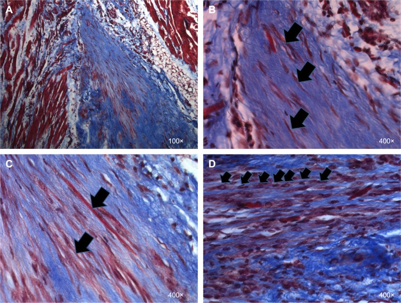
Figure 2.
Evidence for new tissue formation in VML injuries repaired with TEMR–MDC constructs.
Notes: LD muscles repaired with TEMR–MDC (n=10) constructs were retrieved 2 months postimplantation, paraffin embedded and processed for Masson’s trichrome staining, and analyzed for morphology and new tissue formation. Red indicates muscle, blue indicates collagen, and black indicates nuclei in Masson’s trichrome staining. (A) An overview at the interface of native tissue and scaffold. (B) Arrows indicate regenerating striated muscle fibers. (C) Arrows indicate long, differentiated MDCs and neofibers on the scaffold, whereas arrows in (D) indicate fusion of MDCs to form new muscle fibers.
Abbreviations: VML, volumetric muscle loss; TEMR, tissue-engineered muscle repair; MDC, muscle-derived progenitor cell; LD, latissimus dorsi.

