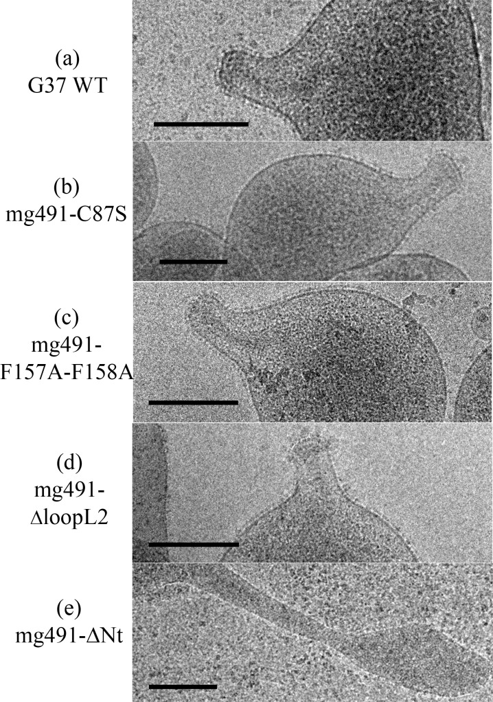
Fig 8. Cryo-electron microscopy analysis of M. genitalium G37 wild type and MG491 mutant strains.
(a) G37 wild type cells showing the cytoskeleton supporting the terminal organelle and the nap layer surrounding the cell membrane. (b) mg491-C87S, (c) mg491-F157A-F158A and (d) mg491-ΔloopL2 cells, exhibiting normal cytoskeletons and nap layers. (e) mg491-ΔNt cells showing no internal terminal organelle structure inside the filament. Bars are 0.2 μm.

