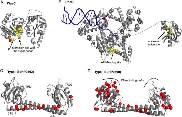
Figure 3.
Amino acid residues under diversifying selection mapped on structural models. Residues under diversifying selection (dN/dS > 1) in H. pylori proteins are shown as red spheres, and those interacting with any ligand in homologous proteins are shown as yellow spheres. (A) WaaC (HP0279). Red sphere, Y229. (B) RecB (HP1553). Red spheres, L680 and R681. (C) HsdS (Type I S subunit) (HP0462). Red spheres, 153, 158, 159, 163, 166, 177, 180, 188, 190, 198, and 346. (D) HsdS (Type I S subunit) (HP0790). The 44 positions shown in red correspond to the codons under diversifying selection.

