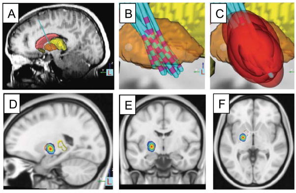
Figure 2.
Deep brain stimulation target in the globus pallidus based on retrospective analysis of the site of effective electrode contacts and modeling of stimulation fields. MRI was used to identify the location of the DBS electrode in patients with DYT-1 dystonia (A) and co-registered into a common atlas space (B). The stimulation field for the effective electrode contact in each patient was modeled (C). A probabilistic volume in the posteroventral aspect of the GPi was identified that could be used to guide future electrode placement or programming (D-F). Modified with permission from Cheung et al. 2014 Annals of Neurology.

