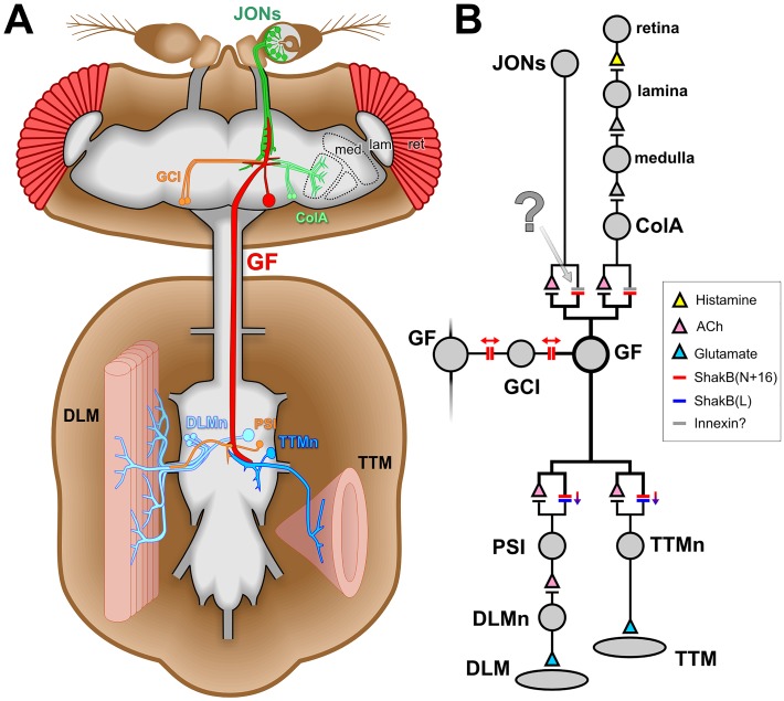
Fig 1. The Drosophila giant fiber escape circuit.
(A) Diagram of the anatomy of one half of the system, it being duplicated contralaterally. In the brain, the giant fiber (GF) receives synaptic inputs onto its dendritic branches from the mechanosensory Johnston’s organ neurons (JONs) and from polysynaptic visual pathways (ret: retina, lam: lamina, med: medulla, ColA: lobular ColA interneurons). It also forms synaptic connections with the giant commissural interneurons (GCI). The GF axon descends to the thoracic ganglion where it forms electrical and chemical synapses with the tergotrochanteral motorneuron (TTMn) of the cylindrical tergotrochanteral (jump) muscle (TTM) and the peripherally-synapsing interneuron (PSI) which innervates the dorsal longitudinal motorneurons (DLMn) of the dorsolongitudinal flight muscles (DLM). (B) Wiring diagram of the circuit. Chemical synapses are denoted by triangles, colored to represent the transmitter where it is known. Electrical synapses are denoted by double bars, colored to represent which innexin is involved, with arrows indicating putative rectifying or non-rectifying junctions. The synapse investigated in the current study is indicated with a question mark. Evidence compiled from several publications [5,7,9–11].

