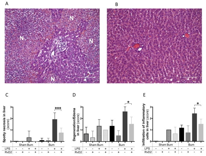
Figure 2. Pathology of the liver after burn injury and endotoxin injection with and without Resolvin D2 treatment.
A. Spotty necrosis of the liver at day 11 pb, after burn and LPS, untreated group (24 hours after LPS injection). Several necrotic foci (N) were identified in the liver tissue sections, in the burn and LPS group. Large numbers of inflammatory cells, including neutrophils, macrophages, and lymphocytes infiltrated the portal area (H&E, ×200, scale bar 100 μm). B. Representative section of liver at day 11 pb, in the burn and LPS with RvD2 treatment groups. Spotty necrotic changes were significantly diminished in the RvD2 treatment group (H&E, ×200, scale bar 100 μm). C – E. Quantitative analysis of pathological changes in the liver (N=3 rats total, N=2–3 experiments for each sham burn group; N=6 rats total, N=3–4 experiments for each burn group). We quantified the spotty necrosis, degeneration or edematous changes of hepatocytes, and infiltration of inflammatory cells in the portal area. RvD2 treatment suppressed these impaired pathological changes in liver (Burn/LPS vs. Burn/LPS/RvD2; *p≤0.05). In particular, RvD2 treatment reduced spotty necrotic changes in hepatocytes (Burn/LPS vs. Burn/LPS/RvD2; ***p≤0.001).

