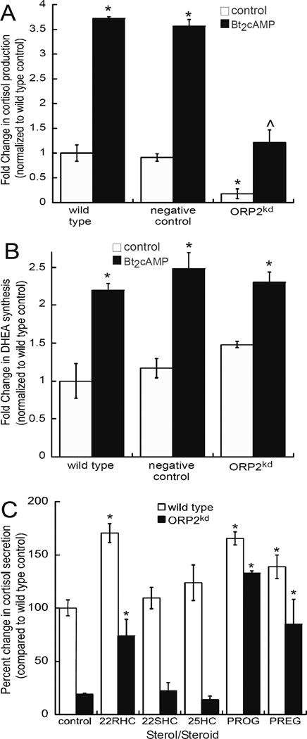
Figure 2. Silencing ORP2 diminishes cortisol secretion.
Cortisol (A) and DHEA (B) concentrations was measured in medium collected from wild type, scrambled shRNA, or ORP2kd H295R cells that were treated for 48 h with 0.4 mM Bt2cAMP by ELISA and normalized to total cellular protein content. ORP2kd hormone levels reflect the average of data obtained from both ORP2 shRNA#1 and ORP2 shRNA#2 clones. Data are graphed as fold change normalized to the metabolite secreted from untreated wild type cells and represent the mean ± SEM of four separate experiments, each performed in triplicate. Asterisks (*) and carats (^) indicate a statistically significant difference (p < 0.05) compared to the wild type control and wild type Bt2cAMP-treated cells, respectively. (C) ELISA assays were used to determine the amount of cortisol secreted into media that was collected from wild type or ORP2kd H295R cells that were treated for 24 h with 10 µM of 22(R)-hydroxycholesterol (22RHC), 22(S)-hydroxycholesterol (22SHC), 25-hydroxycholesterol (25HC), progesterone (PROG) and pregnenolone (PREG). Cortisol amounts were normalized to the concentration of cellular protein and data graphed represent the mean ± STD of three separate experiments, each performed in triplicate. Asterisks denote a statistically significant difference (p < 0.05) when compared to the wild type control cells.

