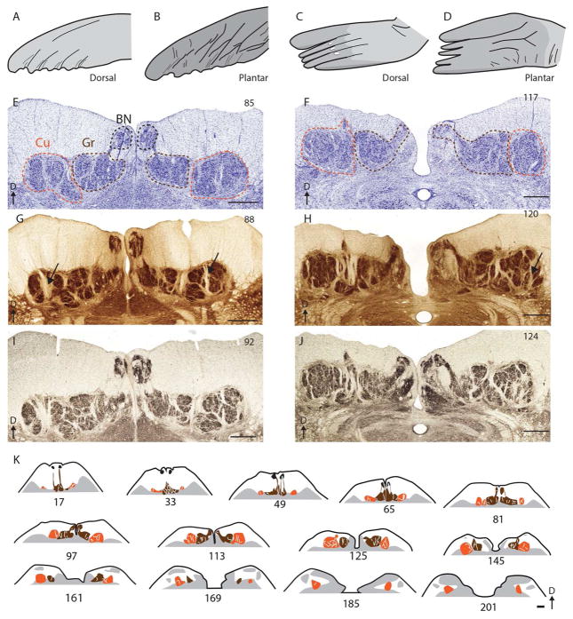
Figure 5.
The dorsal column nuclei (DCN) of the California sea lion. (A) and (B): Sketch of the forelimb of a sea lion. (C) and (D): Sketch of the hindlimb of a sea lion. (E) and (F): Photomicrographs of the DCN stained for Nissl substance. The number of the section is indicated in the upper right. The cuneate, gracile, and Bischoff’s nucleus is outlined in dotted lines of orange, brown and black, respectively. (G) and (H): Photomicrographs of the cuneate-gracile complex stained for cytochrome oxidase, labeled as in A and B. (I) and (J): Photomicrographs of the cuneate-gracile complex stained for vesicular glutamate transporter 1, labeled as in A and B (K): Sketches of the cuneate-gracile complex showing the throughout its rostral caudal extent, from section 17 (most caudal) to 201 (most rostral). The cuneate, gracile, and Bischoff’s nucleus are colored orange, brown and black, respectively. Scale bar = 1 mm. Abbreviations: BN= Bischoff’s nucleus, Cu= cuneate nucleus, D= dorsal, Gr= gracile nucleus.

