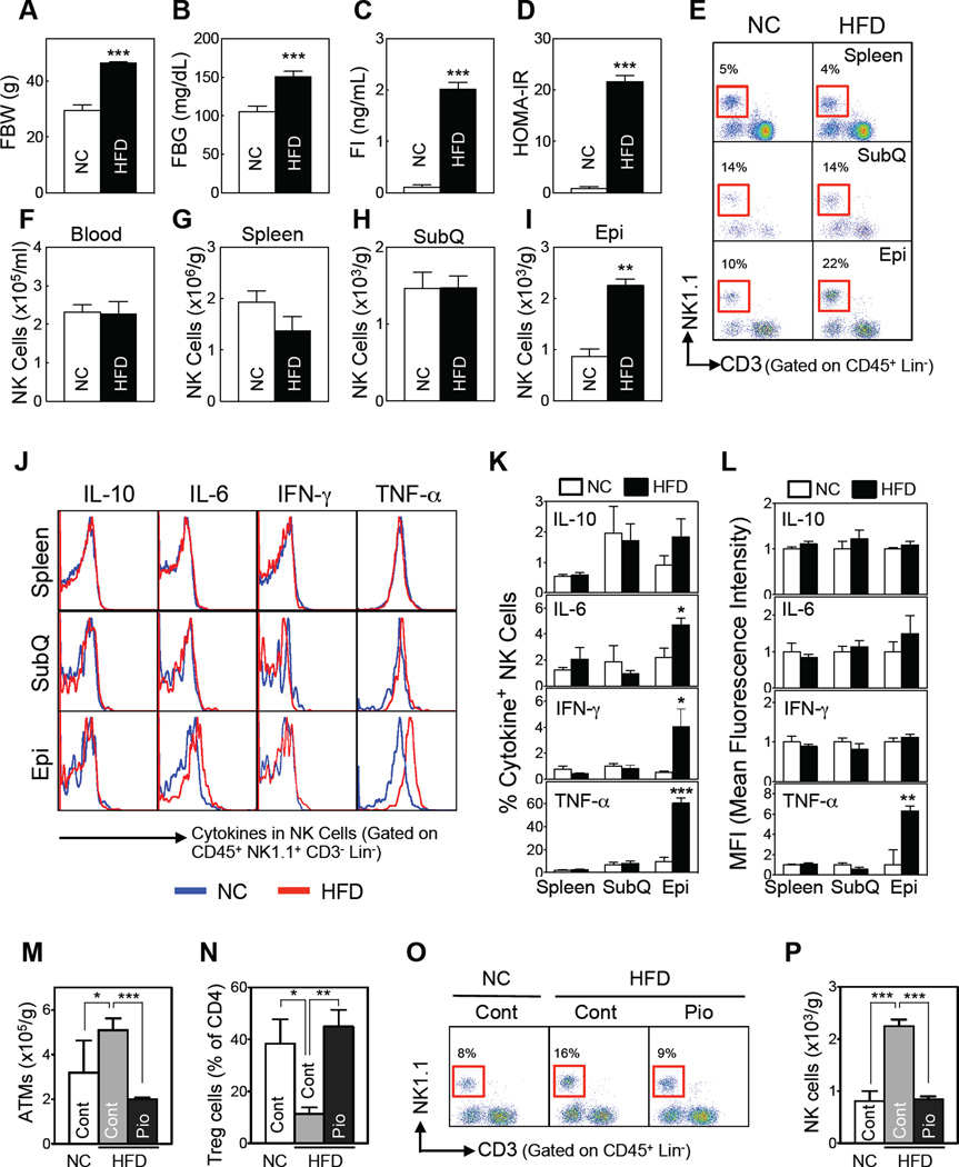
Figure 1. HFD-induced obesity increases adipose tissue NK cell numbers and activation.
(A–L) C57BL/6 male mice were fed a HFD or NC for 12 weeks. (A) Fasting body weight. (B) Fasting blood glucose levels. (C) Fasting serum insulin levels. (D) HOMA-IR (n=5–7/group). (E–L) HFD-induced changes in NK cell phenotypes. (E) Representative flow cytometric plots of the NK cells (red box). The lineage markers (Lin) were TER-119, CD19, and GR-1 for blood; F4/80 was added for the tissue immune cell analyses. (F–I) NK cell numbers (n=3–4/group) in the blood (F), spleen (G), subcutaneous fat (SubQ) (H), and epididymal fat (Epi) (I). (J–L) Flow cytometric analysis of the ex vivo cytokine production by tissue NK cells (n=6/group). (J) Representative cytokine expression. (K) Frequencies of cytokine+ NK cells. (L) Mean fluorescence intensity. (M–P) Pioglitazone (Pio)-induced changes in epididymal fat immune cell numbers. Seven-week-old C57BL/6 mice (n = 8) were simultaneously treated with a HFD (or NC) and Pio (100 mg/kg diet) for 8 weeks. After overnight fasting, epididymal ATMs, Tregs, and NK cells were analyzed by flow cytometry. (M) ATM numbers. (N) Treg numbers. (O) Representative flow cytometry plots of NK cells (red box). (P) NK cell numbers. The data are presented as mean ± S.E.M. *p<0.05, *p<0.01, and ***p<0.001. See also Figure S1.

