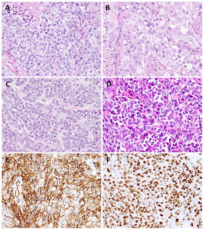
Figure 2. Pathologic features of CIC-rearranged AS.
Solid sheets of monotonous epithelioid cells, with moderate to abundant eosinophilic cytoplasm, and vesicular nuclei (A,B; AS1). Small epithelioid to round cells with scant, light eosinophilic cytoplasm and vesicular nuclei with inconspicuous nucleoli (C; AS2). Distinctive rhabdoid cells arranged in vague pseudoalveolar and nested pattern (D; AS3) All 3 cases expressed diffuse and strong CD31 (E) and ERG (F) immunoreactivity.

