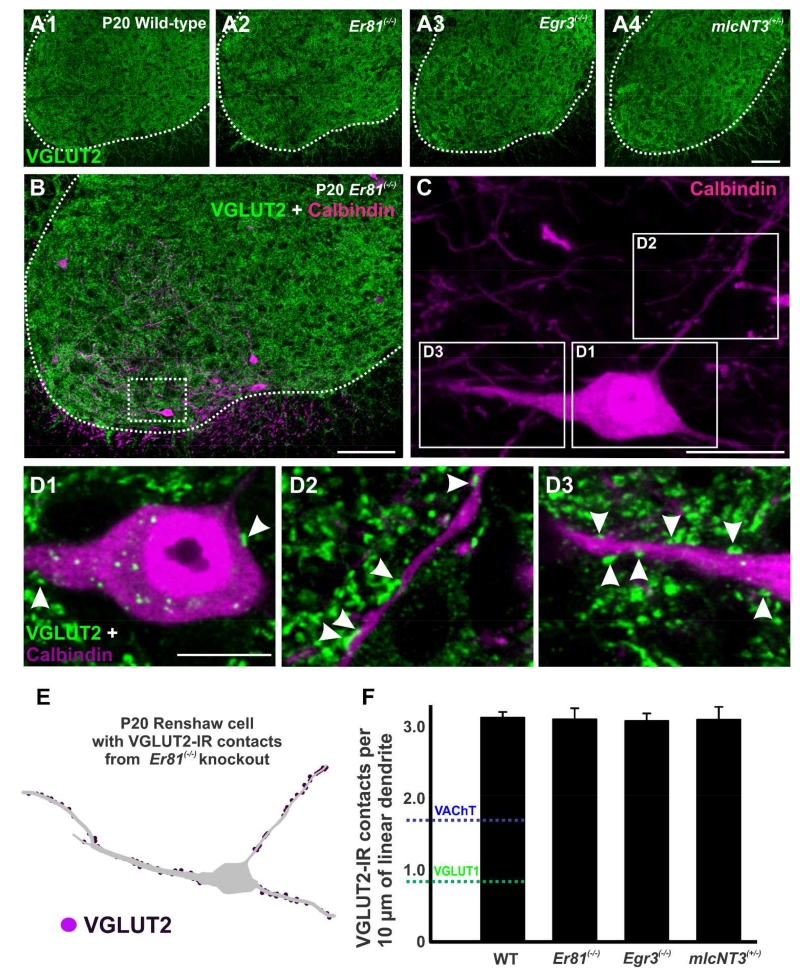
Figure 10. VGLUT2-immunoreactive contacts on Renshaw cells do not change in any mutation.
A, Low magnification confocal images showing VGLUT2-immunoreactivity (green, FITC) in the ventral horn of wild-type (A1), Er81(−/−) (A2), Egr3(−/−) (A3), and mlcNT3(+/−) (A4) mice at P20. The white dotted line represents the border between the ventral horn and the white matter. VGLUT2-IR bouton density is unchanged. B, Confocal image of the ventral horn of a P20 Er81(−/−) spinal cord immunolabeled for CB (magenta, Cy3) and VGLUT2 (green). C, High magnification two-dimensional projection of the CB-IR RC indicated in a box in B. Boxes indicate the dendritic regions shown at higher magnification in D1-D2. D1-D2, High magnification confocal images of VGLUT2-IR contacts (green, arrowheads) on the dendrites of this CB-IR RC (red). E, Reconstruction of this same CB-IR RC showing VGLUT2-IR contacts on the dendrites. F, VGLUT2-IR contact density (per 10 μm of linear dendrite) in P20 wild-type, Er81(−/−), Egr3(−/−), and mlcNT3(+/−)animals. No significant differences were detected in the density of VGLUT2-IR contacts (p=0.997, one-way ANOVA). The blue line represents the density of VAChT-IR contacts and the green line the density of VGLUT1-IR contacts, both on P20 wild-type RCs. Scale bars: 100 μm in A4 and B (A2-A4 are at the same magnification); 20 μm in C; 10 μm in D2 (D1 at the same magnification.

