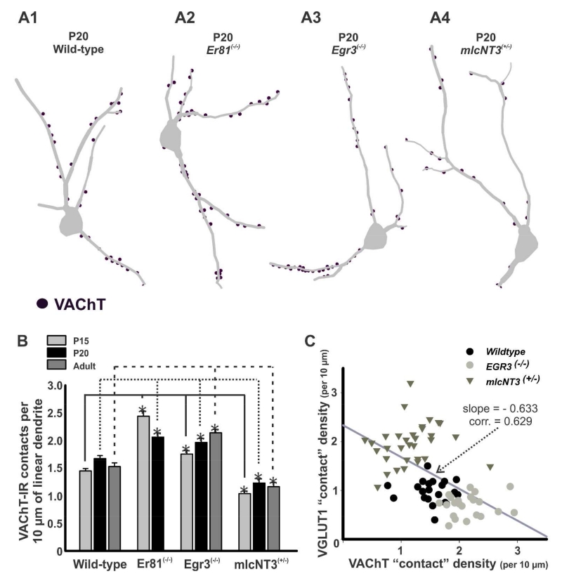
Figure 9. Density of VAChT-immunoreactive contacts on Renshaw cells in wild-type and mutant mice.
A, Three dimensional reconstructions of RCs from P20 wild-type (A1), Er81(−/−) (A2), Egr3(−/−) (A3), and mlcNT3(+/−) (A4) mice. VAChT-IR contacts (dots) are plotted on the traced dendritic arbors. B, Density of VAChT-IR contacts per 10 μm of linear RC dendrite in Er81(−/−), Egr3(−/−), and mlcNT3(+/−) compared to age-matched wild-type controls. During postnatal development RCs from Er81(−/−) animals showed a significant decrease in VAChT-IR contact density from P15 to P20 (p<0.05 in post-hoc Dunn’s comparisons), while it increased in Egr3(−/−) mice from P15 to P20 and adult (p=0.001, one-way ANOVA on Ranks). The density of VAChT-IR contacts was significantly increased at all ages in RCs from Er81(−/−) and Egr3(−/−) animals compared to wild-types (asterisks, p<0.05 in post-hoc Dunn’s comparison). In contrast, VAChT-IR contact densities in mlcNT3(+/−) were significantly lower compared to wild-type animals (asterisks, P15 and P20, p<0.05 in post-hoc Dunn’s comparisons). C, The density of contacts from VGLUT1-IR and VAChT-IR boutons were negatively correlated in a sample population of RCs from preparations in which both synaptic inputs where simultaneously analyzed in single RCs using triple immunofluorescence.

