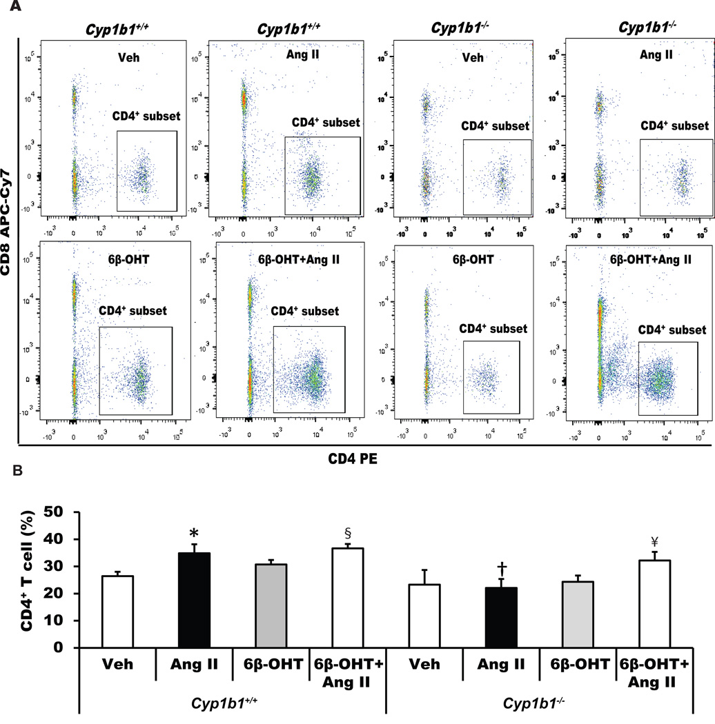
Figure 4. Cyp1b1 gene disruption minimizes angiotensin II (Ang II)-induced renal accumulation of renal CD4+ T-cells in Cyp1b1−/− mice, which is restored by 6β-hydroxytestosterone (6β-OHT).
Cyp1b1+/+ and Cyp1b1−/− mice were infused with vehicle or Ang II and treated with 6β-OHT or 6β-OHT+Ang II for 2 weeks. At the end of Ang II infusion, kidneys were removed and processed, and cells were isolated, counted, and used for flow cytometry. (A) Dot plot images of intact Cyp1b1+/+ and Cyp1b1−/− mice infused with vehicle or Ang II and treated with 6β-OHT for 2 weeks. (B) the average percentage of CD4+ T cell subset is represented by a histogram. *P<0.05 Ang II vs. vehicle; †P<0.05 Cyp1b1−/− Ang II vs. Cyp1b1+/+ Ang II; § Cyp1b1+/+ 6β-OHT Ang II vs Cyp1b1+/+ 6β-OHT; ¥ P<0.05 Cyp1b1−/− 6β-OHT Ang II vs. Cyp1b1−/− 6β-OHT (n=3 for each treatment group and data are expressed as mean±SEM).

