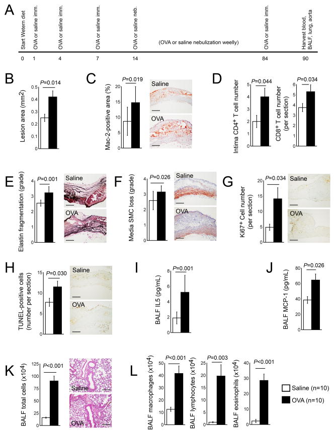
Figure 1.
Chronic ALI promotes atherosclerosis in Apoe−/− mice. A. Experimental protocol. Aortic arch atherosclerotic lesion area (B), Mac-2-positive macrophage contents (C), intima CD4+ T cell and lesion CD8+ T cell numbers (D), media elastin fragmentation grade (E), media SMC loss grade (F), Ki67+ proliferating cell numbers (G), and TUNEL-positive apoptotic cell numbers (H). BALF levels of IL5 (I), MCP-1 (J), total cell numbers (K), and macrophages, lymphocytes, and eosinophils (L). Representative data in panels C, E–H, and K are shown to the right. Scale: 200 μm.

