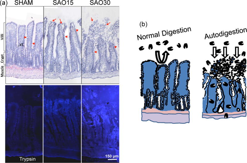
Figure 1.

(a) Example for escape of a digestive enzyme (trypsin) into the wall of the small intestine in the case of splenic artery occlusion (SAO). Top panels show intestinal villi morphology (frozen section labeled with Evans blue) before (Sham) and 15 and 30 min after SAO (SAO15, SAO30, respectively). Bottom panels show corresponding pancreatic trypsin activity (by in-situ zymography according to Methods described in (47)). Bright blue fluorescence indicated cleavage of trypsin specific substrate and is visible inside the villi and across the full thickness of the intestinal wall to the level of the muscle and outer serosa. (Modified from Reference (47)).
(b) Normal Digestion: Schematic illustration of normal containment of digestive enzymes in the lumen of the intestine by the mucosal barrier (mucin layer in conjunction with the epithelium). Autodigestion: entry of digestive enzymes into the wall of the villi and small intestine and destruction of mucosal barrier.
