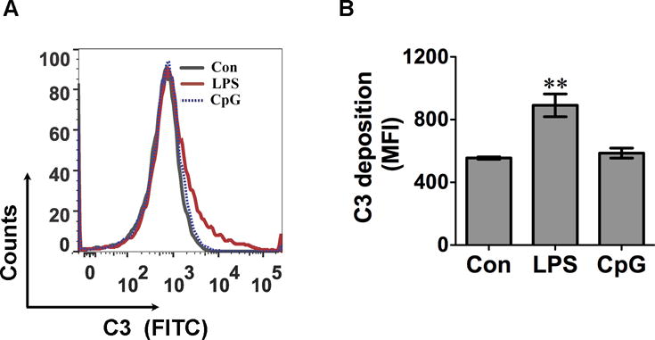
Fig. 2. cfB released from LPS-treated MTECs leads to activation of the complement alternative pathway.

MTECs were stimulated with 500 ng/ml of LPS or 0.25 μM of CpG for 48 hours respectively. Media were collected and incubated with zymosan in the presence of 5 mM of Mg2+, 10 mM of EGTA, and 10% cfB−/− serum at 37 °C for 1 hour. C3 deposition on zymosan was detected by flow cytometry. A. Representative histogram of C3 zymosan deposition. B. Accumulated data of C3 zymosan deposition. The data are presented as mean fluorescence intensity (MFI). N = 3 in each group. **P<0.01 versus control (Con). The experiment was repeated twice. MTEC = mouse tubular epithelial cell, FITC = fluorescein isothiocyanate.
