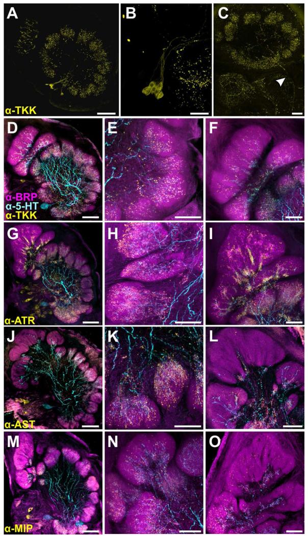
Figure 8.
Distribution of processes from peptidergic neurons and the CSD neurons. A: Tachykinin (TKK) labeling in the AL. Each AL features ~9 TKK-ir cell bodies which in sum innervate all glomeruli B: High resolution scan of TKK-ir cell bodies in the lateral cell cluster. C: TKK-ir process leaving the AL (denoted by white arrow). While most TKK-ir bodies appear to be LNs according to their morphology, the process leaves the AL via the mALT, suggesting that TKK-ir cells may be PNs or centrifugal neurons. D-O: Glomeruli stained for various neuropeptides (yellow), 5-HT (cyan) and BRP (magenta). Left column features the AL (scale bars = 100 um), the middle column features a representative isomorphic glomerulus and the right column features a representative MGC (scale bars = 50 um). D-E: TKK-ir extends father distally than 5HT-ir in isomorphic glomeruli. F: TKK-ir extends farther distally in the MGC than 5-HT-ir. G-I: Dense processes of ATR-ir LNs occur in isomorphic glomeruli and extend farther distally than 5HT-ir. I: Allatotropin (ATR) processes in the MGC extend farther distally than 5-HT-ir. ATR-ir processes are dense proximally but more spare as they extend distally in the MGC. J-L: Allatostatin (AST) processes extend farther distally in the glomeruli than 5HT-ir and are uniform in density throughout. L: AST-ir sparsely innervates the MGC and remains proximal with 5-HT-ir. M-O: Myoinhibitory peptide (MIP) processes extend much farther distally than 5-HT-ir in the isomorphic glomeruli and are uniform in density throughout. O: MIP-ir extends farther distally than 5HT-ir within the MGC.

